The Marvels of Shapes Revealed by Mathematics
The person who I interviewed this time is Dr. Daisuke Ohtsuka, who works in the Laboratory for Developmental Morphogeometry, one laboratory at the BDR that is headed by a team leader with a non-biology background. I thought our conversation might include a lot of references to numbers and mathematical formulas, but as Dr. Ohtsuka himself has a biology background, he enlightened me with a story of when biology and mathematics beautifully intertwine.
Coming to RIKEN made it possible to switch topics
What made you decide to do research at RIKEN?
When I received my doctorate, I was doing research on a virus that infects silkworms that produce silk. The virus is troublesome as the silkworms die when they are infected with this virus. I was in a laboratory in the department of agriculture, so my research was more directly related to the agricultural field. But when I got my degree and began doing my own research, I found myself wanting to do something different; developmental biology was one area I was interested in.
Luckily, there were a few positions that were advertised around that time, one of which was in my current laboratory. My boss was just starting to set up the laboratory at what was then the RIKEN Center for Developmental Biology (CDB), and I was one of the first post-docs hired in the laboratory.
So, I guess you jumped right into a very different field. It must have been quite challenging at first.
Yes, it was. Not only did I switch fields, but my boss and other laboratory members had a background in mathematics, which meant we use different terminology and have different assumed knowledge, so we sometimes had difficulties communicating. I had learned calculus and linear algebra in my classes at university, but had forgotten most of it. So in the early days of joining the laboratory, my co-workers held mathematics tutorials for me, and I, in turn, taught them about biology.
At the time, though, I also did not know a lot about developmental biology. But the CDB was a research institute with a focus on developmental biology, so everyone around me was an expert in this field. I learned about developmental biology by being at the CDB. It was because I came here that I was able to switch fields smoothly.
How shapes are formed
When you switched fields, had you already decided what your project would be?
The fundamental theme of the laboratory is to understand how shapes of organisms are formed. We knew that we would start by adopting the approach taken by my boss, Dr. Yoshihiro Morishita, to analyze limb (arm and leg) formation, that is, to have experimental and theoretical researchers work together to examine the process of shape-changing quantitatively using microscopy data and mathematic formulas. We observe the developmental process of animals using microscopy to track how their shapes change and the cellular phenomena taking place during this process. Where do the cells divide, where do the cells grow, and where do they undergo rearrangement? I was to observe these phenomena and describe the process as a mathematical formula.
Among the different body regions, I have been focusing on the brain.
How does the brain form?
First, a sheet of neural cells is formed, and this then curls into a tubular structure like a straw. The brain arises from this tubular structure.
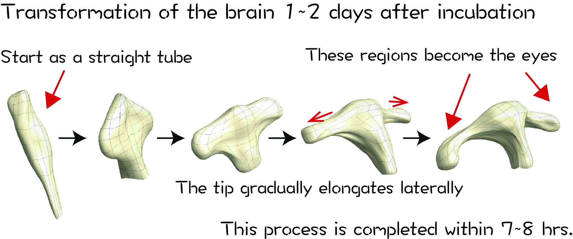
The shape of the tip of the tube is changing, is it not?
That’s right. In humans, the clump of cells at one end of the tube becomes the cerebrum and the section behind the cell clump grows narrower to eventually form the spinal cord. The cell clump at the tip elongates laterally, and this is where the eyes later form.
I am focusing on this area where lateral elongation takes place.
Why do these protrude outward on both sides?
In humans, there is a congenital disease called cyclopia, in which, as implied by the name, results in the formation of only one eye. In most cases, the babies are delivered stillborn, and it is known that a mutation in a particular gene is linked to the onset of this disease. We also know that the activity of this gene leads to the bidirectional protrusions. However, this gene is also known to be involved in the formation of fingers. Without this gene, bone formation would not progress normally and results in only one finger.
The eyes and bones are quite different, though.
Yes, that’s right. The same gene is involved in generating both the eyes and fingers, but we don’t know why this is the case. This was the kind of thing I wanted to know more about, and why I wanted to come here.
How to change the structural shape
How do the tissues elongate laterally? Oh, I guess the cells can just proliferate…?
Actually, it is not that simple (laughing).
What do you mean?
When a structure changes by elongating laterally, there are several possible cellular behaviors to explain this phenomenon.
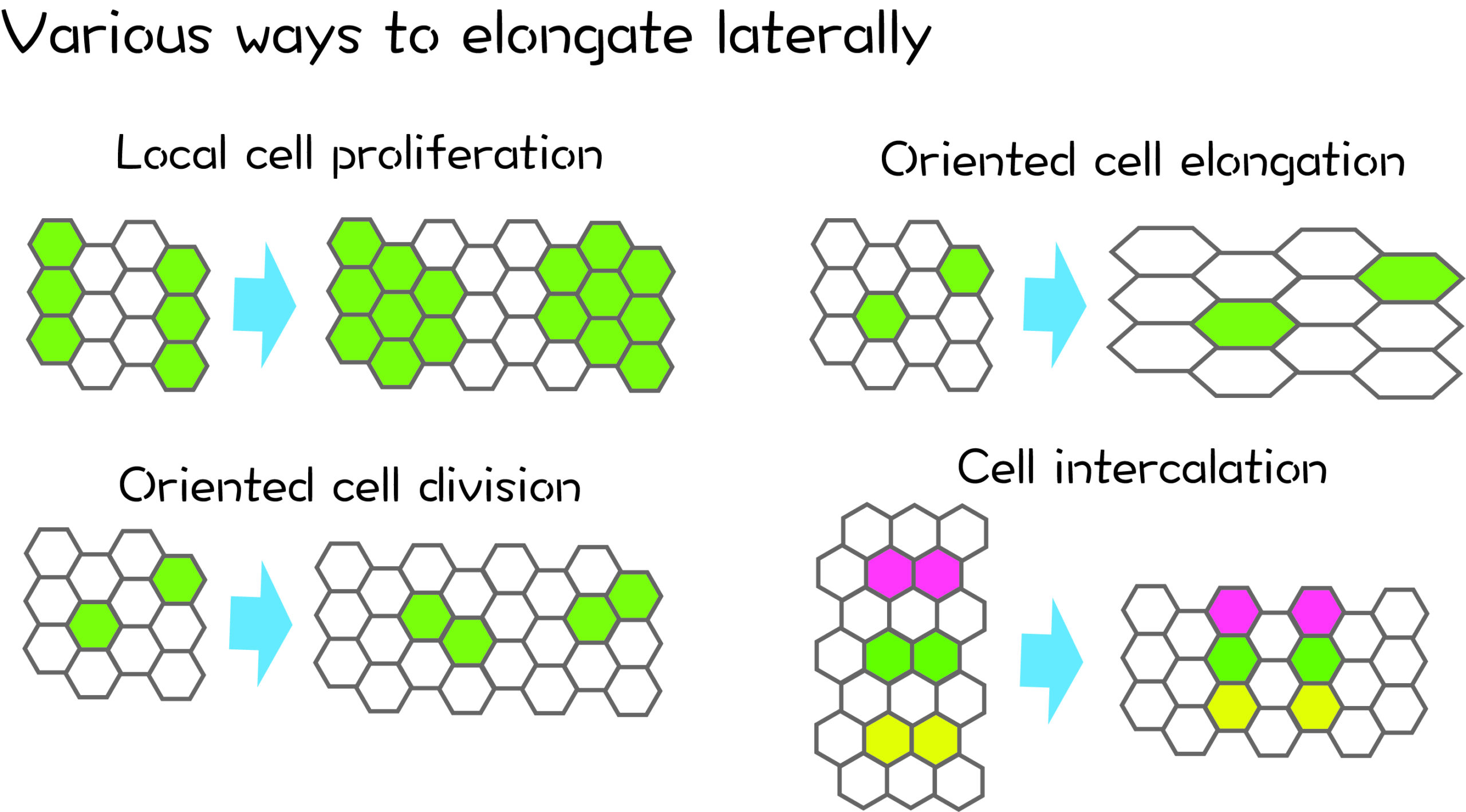
For example, let’s assume that some of the cells are proliferating (upper left panel). Only the green cells are dividing and proliferating, while the white cells hardly increase in number. This would result in the lateral elongation of the tissue overall, right?
I see that the cells in lower left panel have simply doubled in number.
In this case, the cells all divide in the same orientation as the direction of tissue elongation, which leads to the tissue as elongating laterally. Or even if the cells do not proliferate and they simply expand their area in the lateral direction (upper right panel), this would also be an explanation for how the shape of the tissue elongated laterally. In the lower right panel, the cells have changed their alignment, with white cells moving vertically to integrate between the colored cells to change which cells they neighbor. The number of cells does not change, but this too can result in lateral elongation.
So even though you can see that there was an overall change in shape, if you do not closely examine cell behavior and observe how the cells are actually moving, the dynamics of the cells within the tissue remains unclear.
I can certainly see how the tissue (in all the examples) results in lateral elongation.
The gene I mentioned earlier which causes cyclopia is known to stimulate cell proliferation when added to cell culture. Therefore, it was thought that the tissue cannot elongate laterally when this gene is impaired because the cells could not proliferate.
But when we examined the behaviors of cells during the protrusion of the eyes, we discovered that cell proliferation was not involved in this process at all.
I guess you really do need to observe the process to know what is taking place. Are you sure the cells are not proliferating?
We see lateral elongation even if cell proliferation is inhibited. For starters, this elongation process is completed within about seven to eight hours, so that is only enough time for cells to divide once. Even if there was a localized proliferation of cells, it would not have much of an effect on elongation. Therefore, changes in cell alignment have a more significant effect on elongation than an increase in cell numbers.
Under normal conditions, we see cells rearranging to elongate laterally, while this does not occur under abnormal conditions. We looked at what happens when the function of the gene I mentioned earlier is disrupted, and found that direction of elongation becomes randomized, not unidirectional, with the cells displaying disorganized movements within the tissue. And as a result, the tissue does not elongate.
Updating gene annotations
How is the direction determined?
For example, white blood cells which are involved in immunity use concentration gradients to determine the direction of their movement. But in our case, we hypothesized that physical forces were the determinant for how these cells move.
Finally, we talk about forces!
I asked a co-worker in the laboratory, who is an expert in mechanical simulations, to calculate the forces exerted on the tissues, which revealed that the forces are at right angles to the direction of elongation.
But that was just a simulation, right? (sounding unconvinced)
Well, I actually carried out the experiments as well.
(Impressive!)
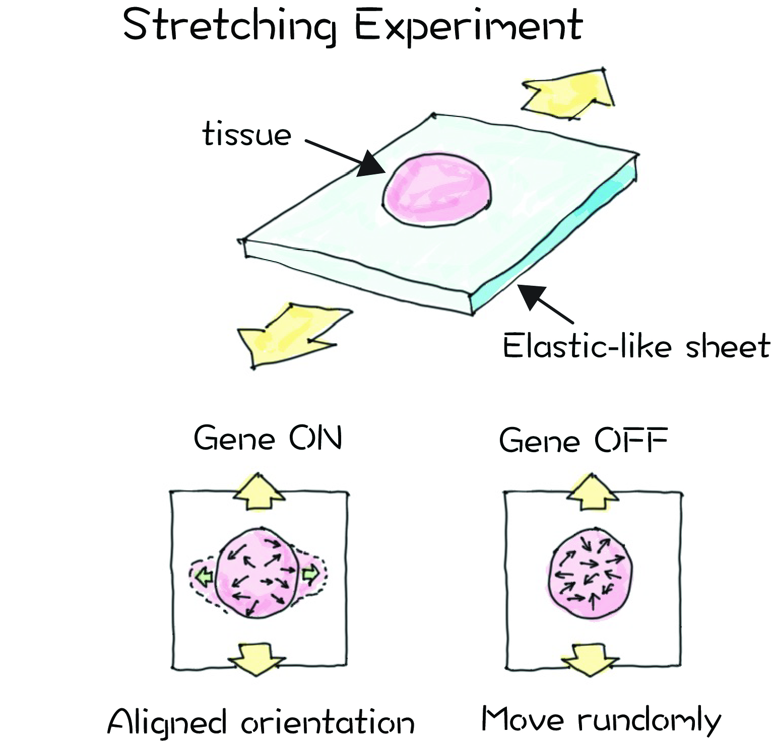
We attached a patch of tissue to an elastic-like sheet and stretch them in longitudinal and uniaxial directions.
It sounds surprisingly like a simple experiment.
Yes, it is. It was a very simple experiment.
If we stretch the patch of tissue in the direction of the force, we see the patch elongating in a direction perpendicular to the that of the force applied, similar to what occurs during elongation of the eyes. When we inhibit the function of the gene described earlier and perform the same experiment, the patch does not elongate even with application of force. This means that the cells cannot sense the stretching force applied to the tissue. In other words, the cells cannot respond to forces without that gene.
Then that gene is like a ‘force sensor’?
Right. This action of cells sensing and responding to force is called ‘mechanosensation’ and the function of this gene is linked to the regulation of this mechanosensory ability. This means that the function of this gene is no longer associated with the bones or brain.
So, the function of that gene is to sense forces and convert that into directional information; not to give commands like, “make a finger”.
I am hoping to reach this kind of generalized understanding of the genes. While we know that loss of function of certain genes leads to the lack of eye formation or finger formation, that is of course the outcome. I don’t think that we have connected all the dots yet on how a gene is involved in the formation of a specific part or how this process can be impaired. That is what I am interested in understanding.
I guess it’s like updating annotations or information on the significance of the gene.
Incidentally, I recently published a new paper reporting on what I just explained about the why in organisms displaying cyclopia, the eyes don’t elongate into two, so please take a look at that as well. We also put out a press release for that paper!
Wow, that work is really hot off the press!
Postscript
When shapes undergo change, I thought that the cells were probably just proliferating, but it’s true that the arrangement of the cells can change. One might think, “Can cells really move that much?” but I guess in fact they do move. It’s oddly curious. Since 2000, we have seen a lot of work on labeling gene functions, and I am sure we will see a lot more advancements in the future. This field seems like it is an extremely exciting one.