Vol.17 – 2024 Winter
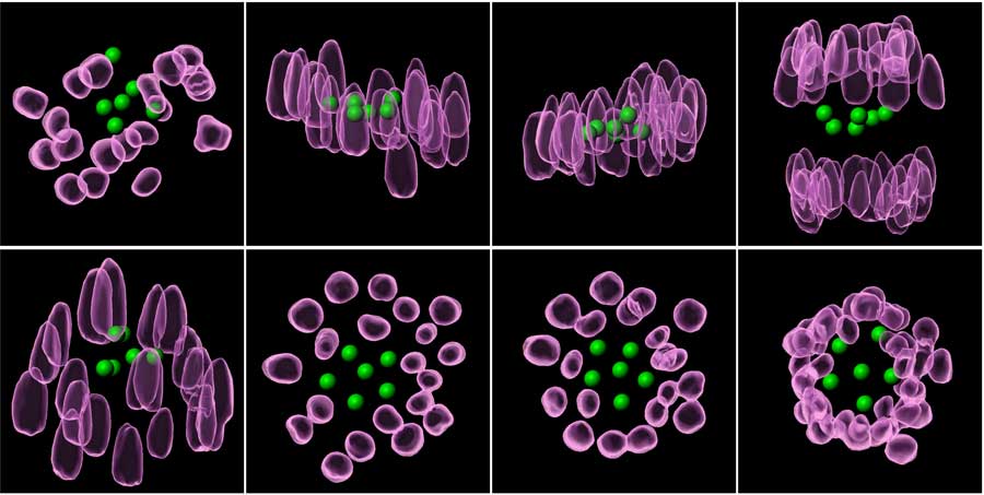
Mysterious stone circle?
These images show the behavior of chromosomes (purple) and artificial kinetochore beads (green) within the cell during metaphase, when they align on the same plane.
Vol.16 – 2024 Summer
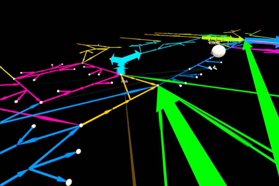
A car navigation screen?
This is a simulation model of the network of chemical reactions (metabolic pathways) occurring within the cell visualized in a virtual reality platform. The metaverse platform allows multiple researchers to analyze and build models collaboratively within a virtual space.
Vol.15 – 2024 Spring
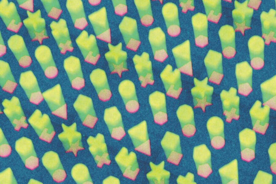
Put the blocks through the holes…
This is an experiment showing the technology allowing scientists to build 3D structures of actin, a protein acting as the cell skeleton. When various subcellular-sized shapes (pink) were drawn on a sheet mimicking the cell membrane (blue), actin (green) was self-assembled into pillars with maintaining the drawn shapes.

Vol.14 – 2023 Autumn
Rain clouds detected by radar?
This is an image of proteins regulating genes in the cell nucleus. In an effort to reveal the behavior of genes in the nucleus of ES cells in two different states, the proteins, which are moving too rapidly to be captured by a camera, are manipulated to fluoresce the moment it binds to DNA.
Vol.13 – Summer 2023
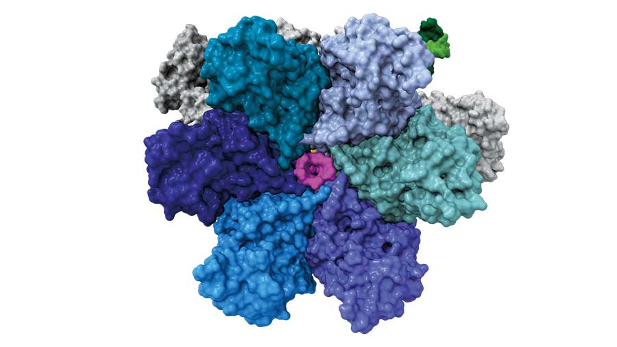
A six-winged pinwheel?
This is the transcription termination factor Rho. It is a ring-shaped structure consisting of a hexamer that forms a tunnel in the center for the RNA (magenta) to pass through. When Rho attaches to the RNA polymerase (white and gray) during transcription, it uses this tunnel to pull the RNA out and terminate transcription at the appropriate time.
Vol.12 – Winter 2023
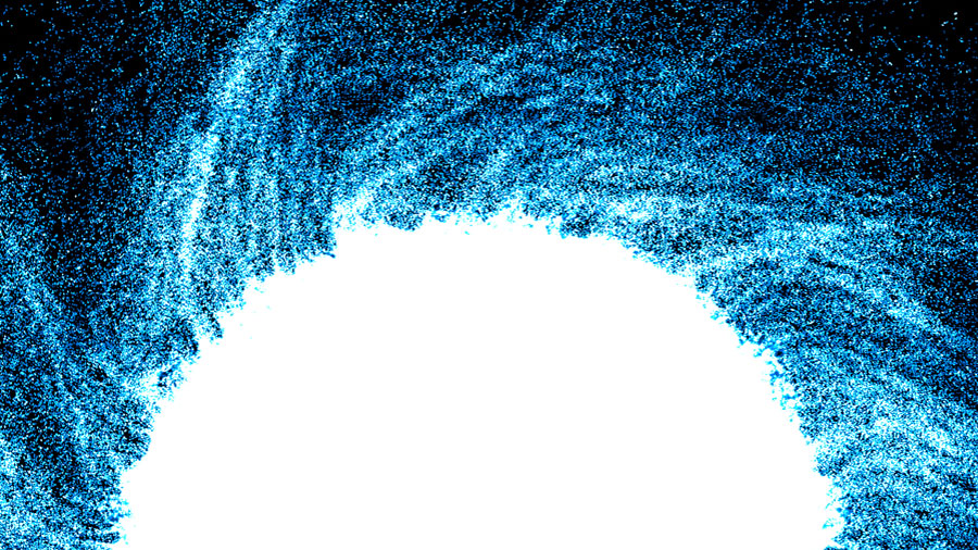
A blue sun?
This is an aggregate of neural stem cells and the outflow of these cells from the aggregate. At high densities, neural stem cells align their elongated shapes and radially crawl out of the aggregate. Their weak clockwise cell motility becomes accentuated at the collective migration level, resulting in the appearance of a spiral formation that is visible to the naked eye.

Vol.11 – Summer 2022
A maze and a cave?
This is a heart of a newborn opossum (red). Opossums are marsupials, like kangaroos. Their hearts retain the ability to regenerate for at least two weeks after birth, which is the longest reported to date in mammals. The green cells are those currently proliferating.
Vol.10 – Spring 2022
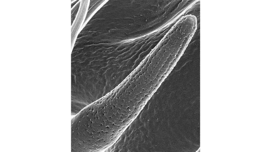
A growing baby corn?
This is an electron microscope image of an olfactory sensillum found on the head of a fruit fly. Nanometer-sized odorant molecules can pass through tiny pore on the sensillum surface to stimulate the nerves inside for detecting odors. In contrast, micrometer-sized dust and virus particles are prevented from entering the body because they cannot pass through the pores.
Vol. 09 – Winter 2022
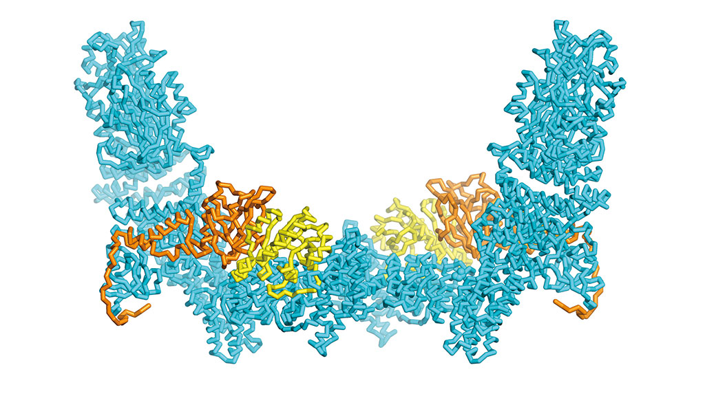
Light blue hammock?
This is a graphic model of the three-dimensional structure of the DOCK-ELMO-Rac protein complex. DOCK (cyan) is involved in cell movement and maturation. The biological activities in the body are maintained by the binding and unbinding of proteins to other proteins.
Vol. 08 – Autumn 2021
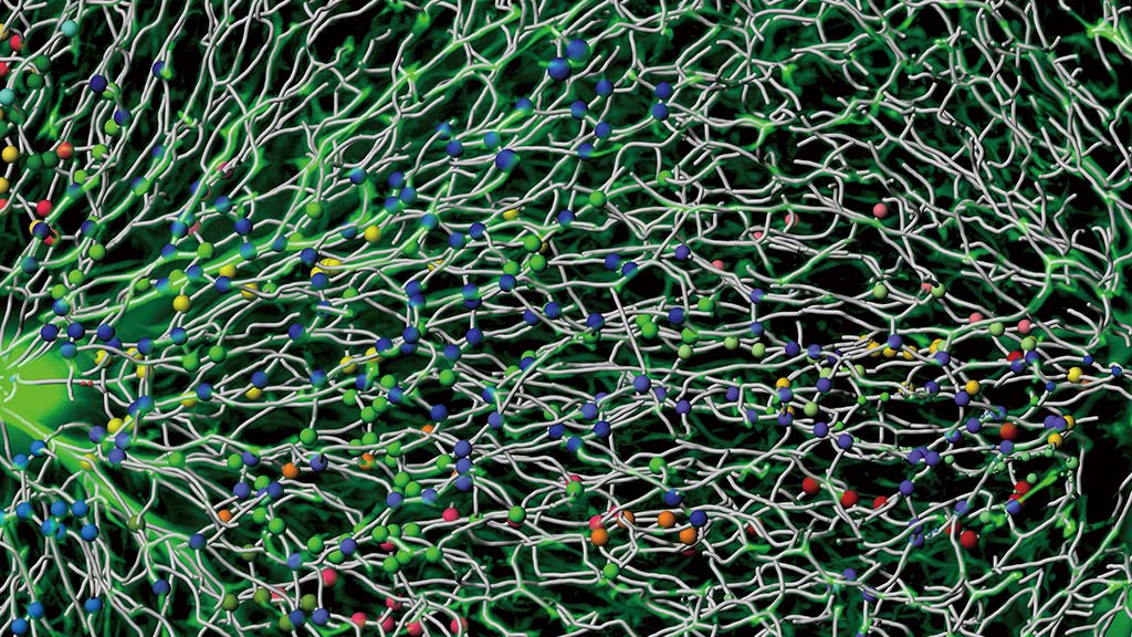
Tangled roots of a tree?
This is a capillary network in the liver called sinusoids. The liver has a high regenerative capacity. It has been shown that when the liver is damaged, there is an increase in blood flow rate and …
Download PDF(1.0MB)
Vol. 07 – Spring 2021
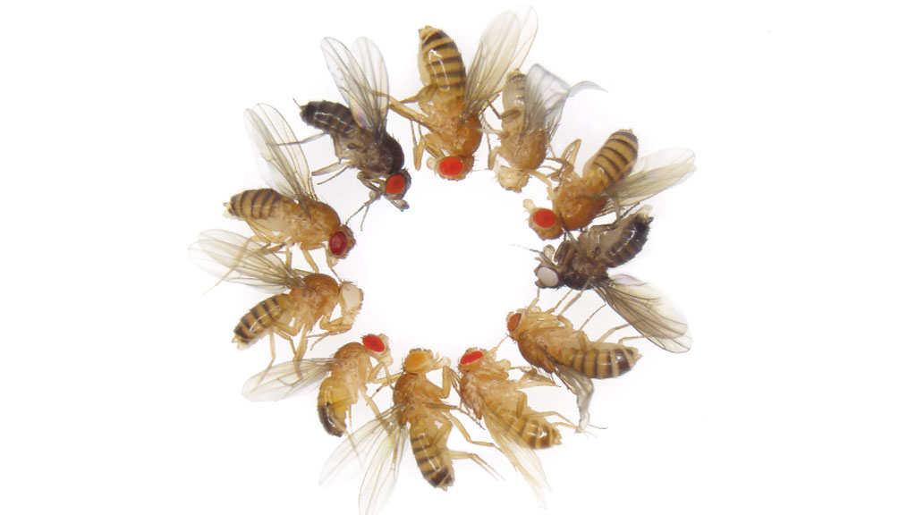
Different fly species?
These are in fact all the same fruit fly species Drosophila melanogaster.
Their different appearances are due to one or multiple genetic mutations, which lead to black bodies, white eyes, etc. There is one wild-type (normal) fly and mutant flies with 20 different phenotypes. Can you identify them all?
Download PDF(536KB)
Read the online version
Vol.06 – Winter 2021
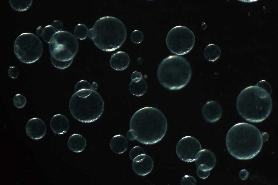
Floating bubbles?
These are 3D alveolar (lung) organoids or mini-organs generated from culturing mouse alveolar stem cells in a culture dish. The organoids can produce different types of alveolar cells.
Download PDF (0.9MB)
Read the online version
Vol.05 – Autumn 2020
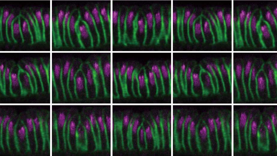
Meadow of flowers in full bloom?
The initiation of cephalic furrow in the Drosophila embryo. This enigmatic structure appears only transiently and yet precisely during development.
Download PDF (0.9MB)
Vol.04 – Spring 2020
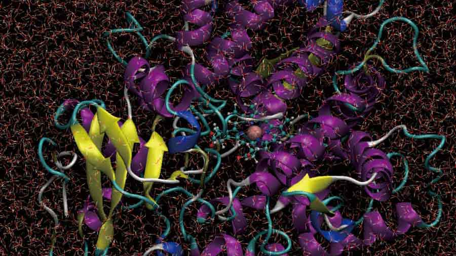
On the cover:
Cytochrome protein containing heme moving in water. Snapshot from a simulation calculated by supercomputer MDGRAPE-4A.
Download PDF (2.8MB)
Vol. 03 – Winter 2019
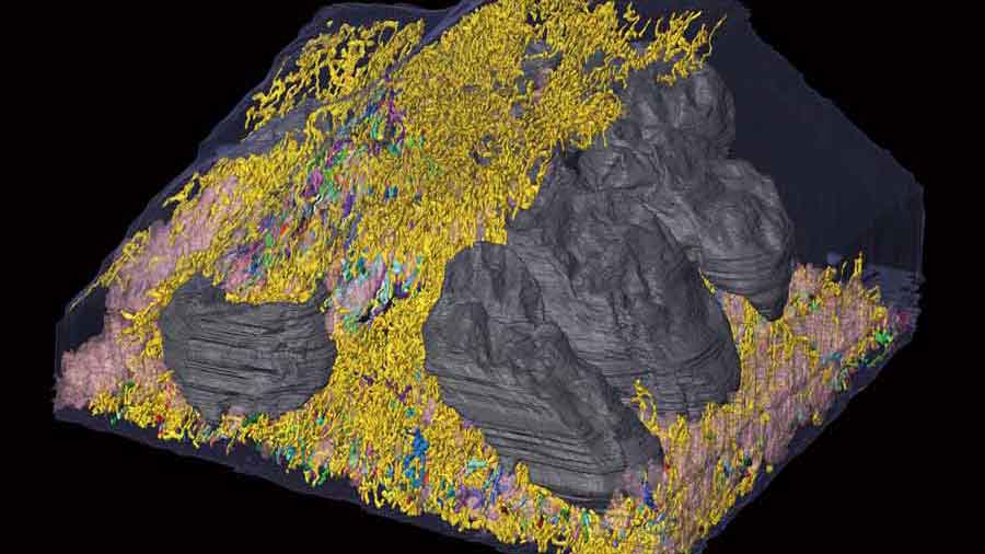
On the cover:
Inside view of a cell captured by electron microscopy. Structure in grey is the nucleus, and structures in yellow and other colors are mitochondria…
Download PDF (1.1MB)
Vol.02 – Summer 2019
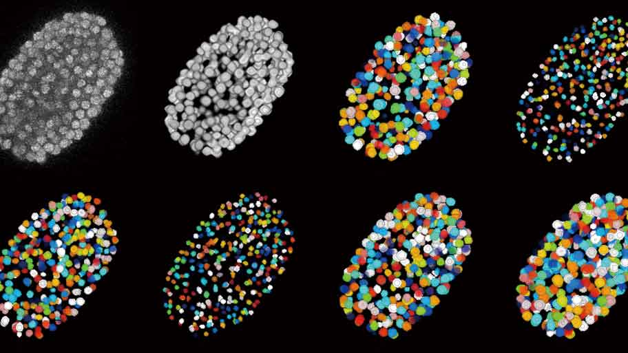
On the cover:
3D microscopic images of C. elegans embryo. Nuclei of the approximately 350 cells in the embryo were labeled with a fluorescent marker …
Download PDF (1.8MB)
Vol.01 – Spring 2019
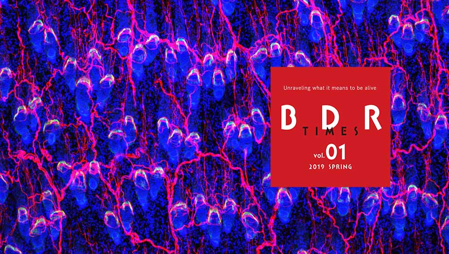
On the cover:
Hair follicle stem cells, which are important for hair shaft generation, secrete extracellular matrix proteins that guide the connections …
Download PDF (2.7MB)