2022-01-14
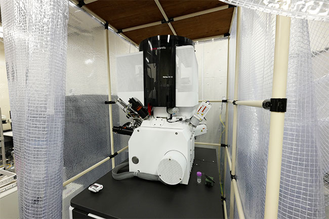
Focused Ion Beam Scanning Electron Microscope
RIKEN–Hiroshima University Collaboration Research Facility
State-of-the-art imaging system protected by two layers of curtains
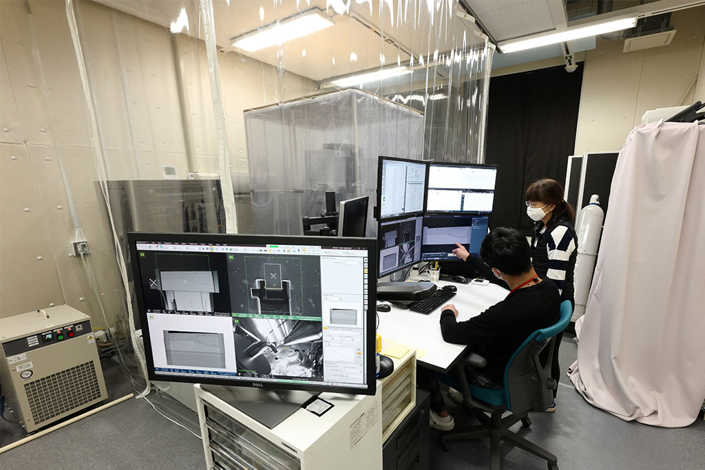
(Top image) The focused ion beam scanning electron microscope (FIB-SEM) is a system which combines focused ion beams for processing or milling the surface of a sample and a scanning electron microscope for observing the surface of a material. Thin, serial, cross-sectional slices are removed from the surface of resin-embedded cell or tissue samples, and then images of each newly revealed layer are captured. The image data are then rendered into 3D using a software program, which then allow scientists to make 3D observations of intracellular structures and their surrounding environment. Operating the instrument is research scientist Takeshi Itabashi and Team Leader Atsuko Iwane (right) of the Laboratory for Cell Field Structure.